|
|
Welcome
to The Heme Protein Database
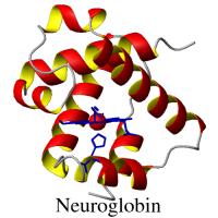
The Heme Protein Database couples
structural information on a
non-redundant set of heme proteins with their associated electrochemical midpoint
reduction potential values. The HPD incorporates the structural data on heme proteins in the RCSB Protein
Data Bank (PDB)
with the protein structure classifications in CATH
3.0.0 and electrochemical data from the primary
literature. The data are presented along with MOLMOL
images of the heme proteins and links to the Prosthetic Groups and Metal Ions in Protein Active Sites Database, PROMISE.
This heme protein structure-function database may be searched in one of the three
ways presented at left. The Search by PDB ID feature allows the user to identify the properties of an individual heme protein.
Data on sets of heme proteins can be retrieved using either the Detailed Search Function,
which yields a list of all the individual members of the set, or the Global View function,
which compiles the data on all the members of the set as a group.
|
|
|
 Detailed
Search
Detailed
Search |
|
Global View |
|